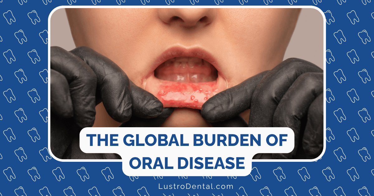
X-ray Types Explained in Plain Language
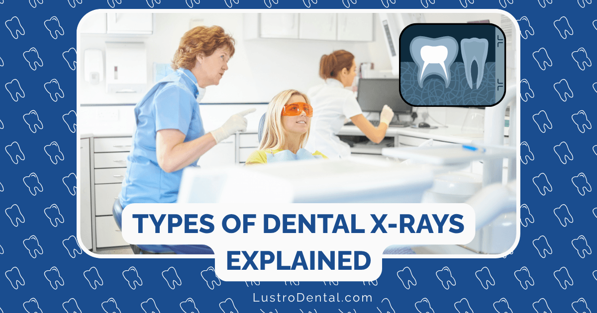
If you’ve ever sat in a dental chair with a heavy lead apron draped over you while a large machine circles your head—or had a small sensor placed between your teeth—you’ve experienced dental X-rays. But do you really understand what these images show and why they’re so important for your oral health? Let’s break down the different types of dental X-rays in simple terms.
Why Dentists Need to See What’s Hidden
Think of your mouth as an iceberg—what your dentist can see during a visual exam is just the tip. Beneath the surface lies a complex world of roots, bone, and developing issues that can only be revealed through X-rays.
According to the American Dental Association, dental X-rays help detect:
- Cavities between teeth that aren’t visible during an exam
- Infections at the root of a tooth
- Bone loss from gum disease
- Impacted teeth (teeth trapped beneath the gums)
- Tumors, cysts, and other abnormalities
Without X-rays, many of these problems would go undetected until they cause pain or visible damage—when they’re typically more difficult and expensive to treat.
The Two Main Categories of Dental X-rays
Dental X-rays fall into two main categories based on where the image receptor (the part that captures the image) is placed:
1. Intraoral X-rays (Inside the Mouth)
These are the most common type of dental X-rays. The image receptor is placed inside your mouth to capture detailed images of individual teeth and the surrounding bone.
2. Extraoral X-rays (Outside the Mouth)
These X-rays are taken with the image receptor outside your mouth. They show broader views of the jaws, skull, and teeth, but with less detail than intraoral X-rays.
Common Types of Intraoral X-rays
Bitewing X-rays
What they show: The crowns (visible portions) of your upper and lower teeth in one area of your mouth.
Named because: You bite down on a wing-shaped holder that positions the X-ray film or sensor.
When they’re used: During routine check-ups to detect:
- Cavities between teeth
- Early signs of bone loss from gum disease
- Changes in the bone density
- Problems with existing fillings
In plain language: Think of bitewings as “cavity detectors” that help catch decay between teeth before it becomes visible or causes pain.
Periapical X-rays
What they show: The entire tooth from the crown to the root tip, including the surrounding bone.
Named because: “Peri” means around, and “apical” refers to the apex or tip of the root.
When they’re used: When you have:
- Pain in a specific tooth
- Signs of infection
- After trauma to evaluate root damage
- Before and after root canal treatment
In plain language: These are “whole tooth” pictures that help your dentist see what’s happening with the entire tooth, including the parts hidden beneath your gums.
Occlusal X-rays
What they show: A broad view of an entire arch of teeth in either the upper or lower jaw.
Named because: “Occlusal” refers to the biting surfaces of teeth.
When they’re used: Most commonly for children to:
- Track the development and placement of teeth
- Detect extra teeth or teeth that haven’t erupted
- Find foreign objects, cysts, or tumors
In plain language: These are “big picture” shots of one jaw that are particularly helpful for monitoring children’s dental development.
Common Types of Extraoral X-rays
Panoramic X-rays
What they show: A single image of your entire mouth, including all teeth, both jaws, temporomandibular joints (TMJs), and sinuses.
Named because: They provide a panoramic or wide view of your entire mouth.
When they’re used:
- As a screening tool for new patients
- To plan for braces, implants, or dentures
- To evaluate wisdom teeth
- To detect jaw problems
- To investigate jaw pain
In plain language: This is the “big picture” X-ray that gives your dentist a comprehensive overview of your entire mouth in one image. You’ll stand still while a machine rotates around your head to capture the image.
Cephalometric X-rays
What they show: A side view of your head that displays the teeth, jaws, and surrounding facial structures.
Named because: “Cephalometric” refers to measurements of the head.
When they’re used:
- By orthodontists to plan treatment
- To analyze the relationship between teeth, jaws, and facial profile
- To track growth and development
In plain language: These are “profile pictures” of your skull that help orthodontists understand how your teeth, jaws, and facial features relate to each other.
Cone Beam Computed Tomography (CBCT)
What they show: Detailed 3D images of your teeth, soft tissues, nerve pathways, and bone.
Named because: The X-ray beam is cone-shaped and rotates around your head to create a 3D image.
When they’re used:
- For complex cases requiring 3D visualization
- Planning dental implant placement
- Evaluating jaw disorders
- Locating the exact position of impacted teeth
- Assessing complex root canal cases
In plain language: This is like a “3D map” of your mouth that gives your dentist extremely detailed information for complex procedures. It’s not routinely used but is invaluable for certain complex treatments.
How Often Should You Get Dental X-rays?
The frequency of dental X-rays depends on your:
- Age
- Current oral health
- Risk for disease
- Signs and symptoms of oral disease
- History of gum disease or tooth decay
According to the Cleveland Clinic, healthy adults with no significant dental issues might need bitewing X-rays every 18-36 months. Those with higher risk factors might need them every 6-18 months.
Children and teenagers often need X-rays more frequently because their teeth and jaws are still developing, and they’re more prone to tooth decay.
Are Dental X-rays Safe?
Many people worry about radiation exposure from dental X-rays. Here’s what you should know:
- Modern dental X-rays use very low doses of radiation
- Digital X-rays (which most dentists now use) reduce radiation exposure by 80-90% compared to traditional film X-rays
- Protective lead aprons and thyroid collars further minimize exposure
- The radiation dose from four bitewing X-rays is comparable to:
- A few days of natural background radiation
- The radiation exposure from a short domestic flight
To put it in perspective, according to Delta Dental, a full mouth series of dental X-rays exposes you to roughly the same amount of radiation you’d receive from daily environmental sources over a few days.
The benefits of detecting dental problems early through X-rays far outweigh the minimal risks associated with the low radiation exposure.
Understanding Your X-ray Results
When your dentist reviews your X-rays, they’re looking for:
- Dark spots on teeth: These often indicate decay (cavities)
- Dark areas around the root tips: May suggest an infection or abscess
- Changes in bone density: Could indicate gum disease
- Position and development of teeth: Important for children and those with wisdom teeth
- Abnormal growths: Such as cysts or tumors (rare but important to detect)
Don’t hesitate to ask your dentist to show you your X-rays and explain what they see. Most dentists are happy to point out areas of concern and help you understand your oral health better.
Questions to Ask About Your Dental X-rays
- Which type of X-ray do I need and why? Understanding the purpose helps you appreciate their value.
- How often should I have X-rays taken? This varies based on your oral health history and current condition.
- Are you using digital X-rays? Digital X-rays use less radiation and provide immediate results.
- Can you show me what you see on my X-rays? This helps you understand your oral health better.
- Are there alternatives if I’m concerned about radiation? In some cases, other diagnostic tools might be options.
The Bottom Line
Dental X-rays are an essential diagnostic tool that helps your dentist provide the best care possible. They reveal problems that aren’t visible during a routine examination, allowing for earlier intervention and often less invasive treatment.
While it’s natural to have questions about radiation exposure, modern dental X-rays are safer than ever before, with minimal radiation doses that pose negligible health risks.
By understanding the different types of dental X-rays and their purposes, you can be a more informed partner in your dental care journey.