🦷 General Care
✨ Treatments
👶 Family Care
💚 Wellness
💡 Resources

🦷 General Care
✨ Treatments
👶 Family Care
💚 Wellness
💡 Resources

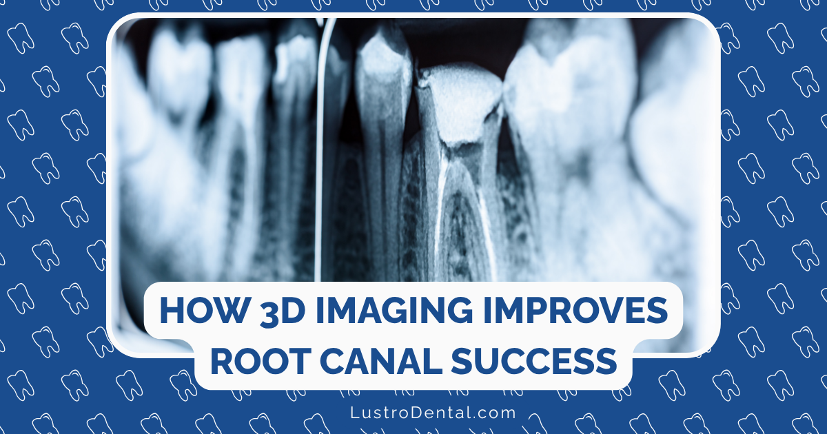
Have you ever wondered why some root canal treatments fail despite the best efforts of skilled endodontists? The answer often lies hidden within the complex anatomy of your tooth—literally. Missing a canal during treatment is one of the most common reasons for root canal failure, and it’s a challenge that has plagued the field of endodontics for decades.
But here’s the good news: advanced 3D imaging technology is changing the game, allowing endodontists to see what was previously invisible and dramatically improving treatment outcomes.
Root canal anatomy is surprisingly complex. While textbooks might show neat, symmetrical canals, the reality is far messier. Teeth can have extra canals, unusual branches, or complex configurations that are nearly impossible to detect with traditional 2D X-rays.
According to a study published in the Journal of Endodontics, up to 40% of treatment failures occur because a canal was missed during the initial procedure. These hidden canals become reservoirs for bacteria, eventually leading to persistent infection and pain.
Dr. Samuel Kratchman, Clinical Professor of Endodontics at the University of Pennsylvania, explains: “The anatomy we can’t see is often what determines our success. Traditional radiography simply doesn’t show us the complete picture.”
Three-dimensional imaging, particularly Cone Beam Computed Tomography (CBCT), has revolutionized how endodontists visualize tooth anatomy. Unlike traditional X-rays that compress three-dimensional structures into flat images, CBCT scanners rotate around the patient’s head, capturing hundreds of detailed images from different angles.
These images are then compiled into a comprehensive 3D model that can be examined from any angle, providing unprecedented detail of the entire root structure.
The CBCT scanning process is remarkably patient-friendly:
Once captured, the endodontist can manipulate the 3D model on a computer screen, examining the tooth slice by slice, rotating it to view difficult angles, and even measuring precise dimensions of canals and surrounding structures.
The impact of 3D imaging on endodontic practice has been profound. Here’s how it’s transforming treatment outcomes:
The maxillary molars (upper back teeth) are notorious for having a fourth canal that’s often missed. Studies using CBCT imaging have shown that the mesiobuccal root of maxillary first molars has a second canal (MB2) in up to 93% of cases—far higher than previously thought.
“Before CBCT, finding the MB2 canal was almost like a treasure hunt,” says Dr. Maria Lopez, an endodontist at Midtown Endodontics. “Now we can pinpoint its exact location before we even begin treatment.”
Vertical root fractures are notoriously difficult to diagnose with conventional imaging but are clearly visible on CBCT scans. Early detection can save patients from undergoing unnecessary treatment on teeth that ultimately need extraction.
Internal and external resorption (breakdown of tooth structure) can be precisely mapped with 3D imaging, allowing for targeted treatment. Similarly, periapical lesions (infections at the root tip) can be detected earlier and their true extent assessed more accurately.
For cases requiring endodontic surgery, CBCT provides crucial information about the proximity of vital structures like nerves, sinuses, and blood vessels. This allows for more precise surgical planning and reduced risk of complications.
Dr. James Chen, an endodontist at Irvine Endodontics, shares a telling case: “A patient came to me after a failed root canal. She’d been in pain for months despite multiple treatments. Her previous 2D X-rays showed nothing unusual, but our CBCT scan revealed a completely untreated canal hiding behind a calcification. We were able to locate and treat it precisely, and her symptoms resolved completely.”
These success stories are becoming increasingly common as more practices adopt 3D imaging technology.
The newest advancement in endodontic imaging combines 3D CBCT technology with artificial intelligence. A single CBCT scan can contain hundreds to thousands of image slices—far more data than a human can efficiently process.
AI algorithms are now being developed to automatically detect anomalies, measure canal dimensions, and even suggest treatment approaches based on similar cases. According to Overjet, a dental AI company, these systems can:
This AI integration not only improves accuracy but also significantly reduces the time required to analyze complex scans, enabling faster diagnosis and treatment planning.
The advantages of 3D imaging extend beyond improved clinical outcomes:
“One of the most valuable aspects of 3D imaging is showing patients exactly what’s happening inside their tooth,” explains Dr. Sarah Johnson of Waterloo West Dental. “When patients can visualize the problem, they better understand the treatment approach and tend to be more cooperative with recommendations.”
By ensuring all canals are located and treated initially, 3D imaging significantly reduces the need for retreatment procedures, saving patients time, money, and discomfort.
The precise visualization offered by CBCT allows endodontists to preserve more healthy tooth structure while still addressing all infected areas—a cornerstone of modern minimally invasive endodontics.
While the benefits are clear, endodontists typically use CBCT selectively rather than routinely. Cases that particularly benefit from 3D imaging include:
The American Association of Endodontists has published guidelines for the appropriate use of CBCT in endodontics, emphasizing the principle of ALARA (As Low As Reasonably Achievable) when it comes to radiation exposure.
A common question about 3D imaging relates to radiation exposure. While CBCT does involve more radiation than a standard dental X-ray, the doses are significantly lower than medical CT scans and continue to decrease as technology improves.
Modern CBCT units can produce high-quality images with radiation doses as low as 5-80 microsieverts, comparable to just a few days of natural background radiation. Many units also allow for focused “small field of view” scans that target only the area of interest, further reducing exposure.
Dr. Robert White, radiation safety officer at a major dental school, puts it in perspective: “The diagnostic benefits of a properly prescribed CBCT scan far outweigh the minimal radiation risks, especially when the alternative might be a failed procedure or missed diagnosis.”
The integration of 3D imaging in endodontic practice continues to evolve. Emerging trends include:
Similar to GPS navigation, these systems use the 3D scan to guide instruments in real-time during treatment, ensuring precise access to difficult canals.
Some practices are now 3D printing physical models based on CBCT scans for treatment planning and patient education.
Experimental systems overlay the 3D scan information onto the clinician’s field of view during treatment through special glasses or displays.
According to recent research, these technologies are expected to become standard in endodontic practices by 2025-2030, further improving the precision and predictability of root canal treatment.
If you need root canal treatment, especially for a complex case or retreatment, consider asking potential providers about their imaging capabilities. Questions to ask include:
Many endodontists proudly showcase their advanced technology on their websites and social media, making it easier to find those who have embraced these innovations.
The days of “feeling your way” through complex root canal anatomy are rapidly fading as 3D imaging becomes the standard of care in endodontics. By revealing what was previously hidden, this technology is transforming root canal treatment from an often-dreaded procedure with uncertain outcomes to a precise, predictable solution for saving natural teeth.
As Dr. Chen aptly puts it, “3D imaging doesn’t just help us see better—it fundamentally changes what’s possible in endodontic treatment. The hidden canals that once caused so many failures are now routinely found and treated, leading to better outcomes and happier patients.”
For patients, this means fewer failed treatments, less discomfort, and more teeth saved—a true win-win for modern dental care.
Have you had a root canal procedure that utilized 3D imaging? We’d love to hear about your experience in the comments below!