🦷 General Care
✨ Treatments
👶 Family Care
💚 Wellness
💡 Resources

🦷 General Care
✨ Treatments
👶 Family Care
💚 Wellness
💡 Resources

When a root canal treatment fails, the decision to pursue retreatment often comes with questions about the likelihood of success. After all, if the initial treatment didn’t resolve the problem, what guarantee is there that a second attempt will yield better results? The answer often lies in advanced technology that wasn’t necessarily available or utilized during the first procedure—specifically, the dental operating microscope (DOM).
Microscopic endodontics represents one of the most significant advancements in modern endodontic practice, transforming what was once considered an art reliant on tactile sensation into a precise, visually-guided science. This evolution has been particularly impactful in retreatment cases, where success depends on identifying and addressing factors that contributed to the initial failure.
Endodontic treatment has always been challenging due to the microscopic nature of root canal systems and their limited accessibility. For decades, endodontists relied primarily on:
The introduction of magnification has dramatically changed this approach. According to a study published in the Journal of Dentistry, the use of microscopes in endodontic practice has increased from 52% in 1999 to over 90% in recent years. This shift represents more than just adopting new technology—it’s a fundamental change in how endodontic procedures are performed.
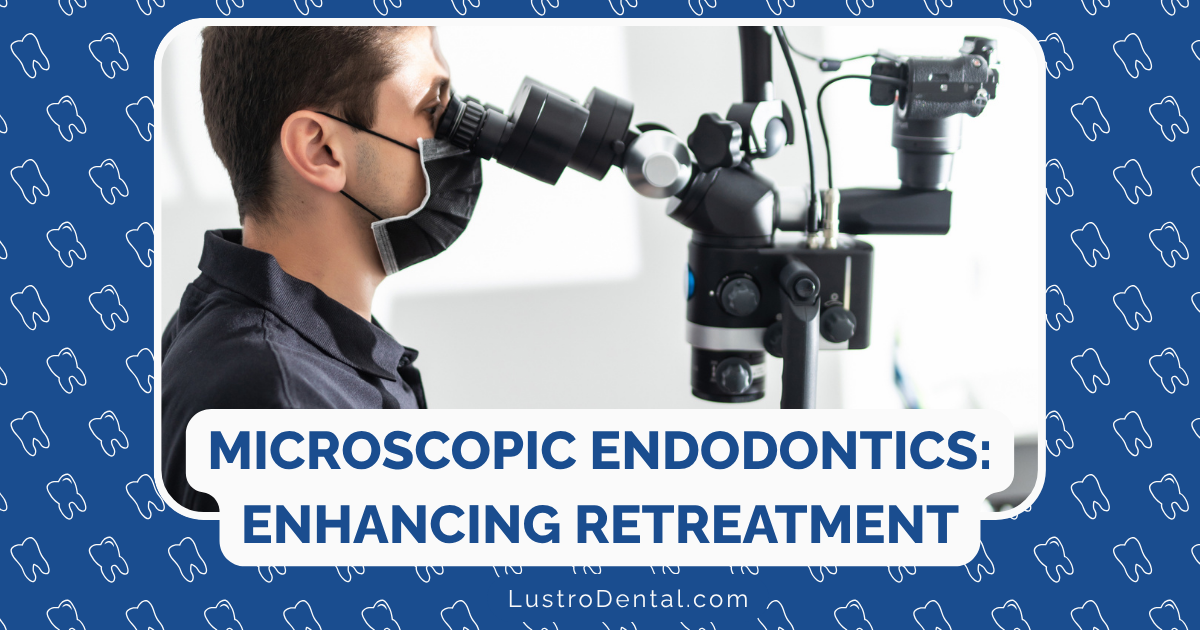
The dental operating microscope provides magnification ranging from 4× to 25× or higher, coupled with coaxial illumination that eliminates shadows in the operating field. Modern microscopes also feature:
Dr. Sarah Johnson, endodontist at Global Surgical, explains: “The microscope transforms what we can see and, consequently, what we can treat. Details that were previously invisible become clearly apparent, allowing for more precise and comprehensive treatment.”
Retreatment cases present unique challenges that make microscopic visualization especially valuable:
Before effective retreatment can be performed, the endodontist must determine why the initial treatment failed. Common causes include:
The microscope allows for detailed examination of the pulp chamber floor and canal orifices, revealing subtle clues that might be invisible to the naked eye.
Root canal systems can be incredibly complex, with lateral canals, isthmuses, and anatomical variations that are difficult to detect without magnification. A 2022 study found that 40 root canal orifices were not detected with the naked eye, but 33 were successfully located using the DOM—an 82.5% success rate for identifying missed canals.
Dr. Michael Chen, researcher in endodontic microsurgery, notes: “The microscope doesn’t just make things bigger; it reveals an entirely new level of anatomical detail that changes our understanding of what we’re treating.”
Retreatment often involves technically demanding procedures such as:
Each of these challenges becomes significantly more manageable with microscopic visualization.
Missed canals are a leading cause of endodontic failure. The maxillary first molar, for example, often has a second mesiobuccal canal (MB2) that can be difficult to locate. Studies have shown:
According to research published in the Journal of Endodontics, teeth treated without microscopes were significantly more likely to have missed canals requiring retreatment.
Separated instruments present a significant challenge in retreatment cases. The microscope provides several advantages:
A clinical study reported in PMC found a 72.3% success rate for removing separated instruments using the microscope combined with ultrasonic techniques.
Complete removal of existing filling materials is essential for retreatment success. Microscopic visualization allows the endodontist to:
Perforations—whether from the initial treatment or created during retreatment—require accurate diagnosis and precise repair. The microscope enables:
The previously mentioned study reported a success rate of 72.7% for managing canal perforations using the DOM, with even higher rates (81.3%) for coronal perforations.
Calcified canals that couldn’t be negotiated during the initial treatment become more manageable with microscopic assistance:
The evidence for improved outcomes with microscopic endodontics is compelling:
A 2025 study in the Journal of Dentistry found that microscope-assisted root canal treatment resulted in:
Overall success rates for endodontic retreatment range from 80-88% according to Endodontic Practice US, with microscope use being a key factor in achieving the higher end of this range.
The impact of microscopes on surgical endodontics has been even more dramatic:
A systematic review published in the International Endodontic Journal found that microsurgical techniques had a periapical healing rate of 94% compared to 59% for traditional approaches.
Different phases of retreatment benefit from specific magnification levels:
Dr. Lisa Wong, endodontic microsurgery specialist, advises: “Learning to work with the microscope is like learning a new language. You start with basic phrases and gradually become fluent. The key is understanding which magnification level serves each specific task best.”
Proper ergonomics is essential for effective microscope use:
A 2025 survey found that improved vision (61.7%) and high precision (35%) were cited as the primary advantages of using magnification systems in endodontic treatments.
Modern microscopic endodontics often integrates with complementary technologies:
This integration creates a comprehensive approach to complex retreatment cases.
Despite its benefits, microscopic endodontics has some challenges:
Mastering the microscope requires time and practice. Many endodontists report:
The financial investment can be substantial:
However, according to the 2025 survey, 40% of practitioners reported recouping these costs within 1-2 years.
Some clinical situations present challenges even with microscopic assistance:
From the patient perspective, treatment by an endodontist using a microscope may differ in several ways:
Dr. Robert Wilson, endodontist, shares: “When I explain to patients how the microscope helps me see and treat problems that might otherwise be missed, they understand the value. Many are fascinated by the technology and appreciate the thoroughness it enables.”
As technology continues to advance, several trends are emerging:
AI systems that can analyze microscopic images in real-time may soon:
Beyond traditional microscopes, new visualization tools include:
As technology evolves, we can expect:
The evidence is clear: microscopic endodontics significantly improves retreatment outcomes. By enabling practitioners to see what was previously invisible, the dental operating microscope has transformed endodontic retreatment from an uncertain proposition to a highly predictable procedure with success rates approaching those of initial treatment.
For patients facing the prospect of endodontic retreatment, seeking a provider who utilizes microscopic techniques can make a substantial difference in the likelihood of success. The microscope doesn’t just magnify the operating field—it magnifies the possibilities for saving teeth that might otherwise be lost.
As one endodontist aptly put it: “Treating root canals without a microscope is like trying to thread a needle in the dark. You might succeed through skill and luck, but why not turn on the light?”
Have you experienced endodontic retreatment with microscopic techniques? Share your thoughts in the comments below.