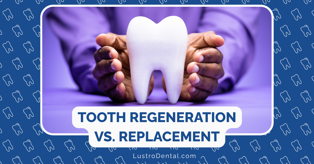
Bioprinting on the Horizon: The Future of Tissue Regeneration in Dentistry
Imagine a future where, instead of receiving a dental implant, your dentist simply prints you a new tooth—a living, biological tooth that integrates perfectly with your body, complete with pulp, dentin, enamel, and a healthy root system. Or picture a scenario where damaged gum tissue is repaired not with grafts, but with customized, living tissue created specifically for you using your own cells.
This isn’t science fiction—it’s the emerging field of dental bioprinting, and it represents one of the most exciting frontiers in dental medicine today. As someone who has followed the evolution of dental technology for years, I can tell you that bioprinting stands to revolutionize how we approach tissue regeneration in dentistry, potentially transforming treatment options for millions of patients worldwide.
In this article, we’ll explore the cutting-edge world of dental bioprinting, examine current research and breakthroughs, and look ahead to what the future might hold for this transformative technology.
Understanding Bioprinting: Beyond Traditional 3D Printing
What Is Bioprinting?
Before diving into its dental applications, let’s clarify what bioprinting actually is:
Bioprinting is a specialized form of 3D printing that uses “bioinks”—materials containing living cells, growth factors, and biocompatible materials—to create tissue structures layer by layer. Unlike conventional 3D printing, which typically uses plastics or metals, bioprinting aims to create functional, living tissues.
According to Nota 3D, the bioprinting process involves:
- Creating a digital blueprint from high-resolution imaging technologies like MRI or CT scans
- Preparing bioinks that maintain cell viability and promote growth
- Printing the tissue construct using specialized techniques
- Maturing the printed tissue in a bioreactor that simulates physiological conditions
The Difference Between 3D Printing and Bioprinting in Dentistry
While traditional 3D printing has already made significant inroads in dentistry (creating crowns, bridges, surgical guides, etc.), bioprinting takes the technology to an entirely new level:
| Traditional 3D Printing in Dentistry | Bioprinting in Dentistry |
| Uses inert materials (resins, ceramics, metals) | Uses living cells and biocompatible materials |
| Creates non-living dental appliances and models | Creates living tissue constructs |
| Primarily for restorative and prosthetic applications | Focuses on regenerative applications |
| End products replace natural structures | End products aim to become integrated, living tissues |
As noted in a comprehensive review published in PMC, this fundamental difference makes bioprinting a potentially transformative approach for treating conditions that currently have limited regenerative options.
Current Applications and Research in Dental Bioprinting
Periodontal Tissue Regeneration
One of the most promising areas for dental bioprinting is in treating periodontal disease—a condition affecting nearly half of adults over 30 in the United States.
The Challenge of Periodontal Disease
Periodontal disease leads to the progressive destruction of the tissues supporting teeth, including:
- Gingival tissue (gums)
- Periodontal ligament (PDL)
- Cementum
- Alveolar bone
Traditional treatments often struggle to fully regenerate these complex, interconnected tissues. However, according to research published in Cell, bioprinting offers a promising solution by enabling the creation of personalized tissue substitutes that can promote regeneration in defective areas.
Current Research Highlights
Recent studies have demonstrated several successful approaches:
- Multi-layered constructs: Researchers have created bioprinted scaffolds with distinct layers mimicking the natural architecture of periodontal tissues
- Cell-laden bioinks: Periodontal ligament stem cells (PDLSCs) incorporated into bioinks have shown the ability to differentiate into the various cell types needed for periodontal regeneration
- Growth factor delivery: Bioprinted constructs can be designed to release growth factors in a controlled manner, enhancing tissue regeneration
A 2025 study cited in PubMed found that bioprinted periodontal constructs showed promising results in preclinical models, with evidence of functional tissue integration and regeneration.
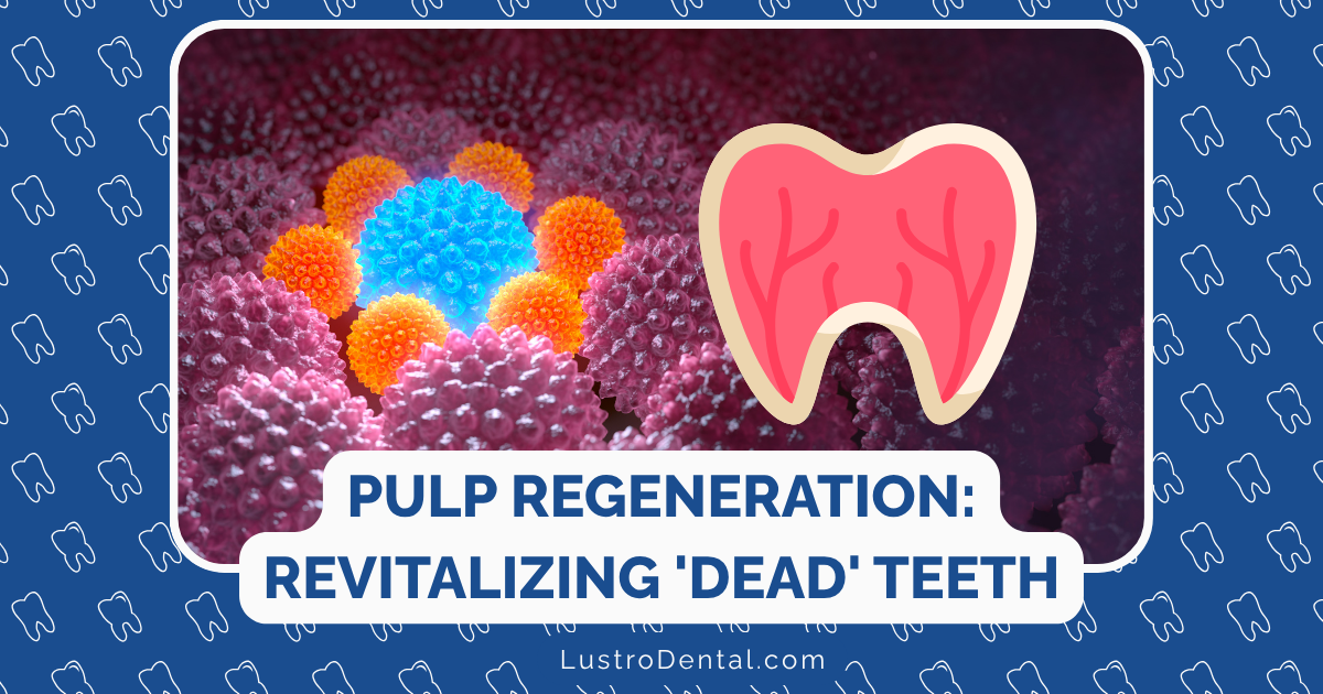
Dental Pulp Regeneration
Another exciting application is in endodontics, particularly for dental pulp regeneration after root canal procedures.
The Limitations of Traditional Root Canals
Traditional root canal therapy involves removing infected pulp tissue and replacing it with an inert filling material. While this addresses infection, it leaves the tooth devitalized—without the nerves, blood vessels, and cells that maintain its health.
Bioprinting Solutions
According to research published in PMC, scientists have successfully bioprinted dental pulp constructs using:
- Dental pulp stem cells (DPSCs)
- Growth factors that promote pulp tissue development
- Biocompatible hydrogels that mimic the natural pulp environment
These constructs have demonstrated the ability to form vascularized pulp-like tissues in preclinical models, potentially offering a pathway to truly regenerative endodontic treatments.
Whole Tooth Regeneration
Perhaps the most ambitious application of bioprinting in dentistry is the regeneration of entire teeth—a goal that would revolutionize how we address tooth loss.
Current Approaches
According to Essential Endodontics, several approaches are being explored:
- Tooth bud bioprinting: Creating artificial tooth buds that, when implanted, develop into functional teeth
- Stem cell-seeded scaffolds: Using bioprinted scaffolds seeded with dental stem cells to guide tooth development
- Gene therapy combined with bioprinting: Activating genes responsible for tooth development in bioprinted constructs
A groundbreaking study reported by Decisions in Dentistry demonstrated the successful creation of bioengineered tooth buds using dental stem cells and decellularized tooth bud extracellular matrix scaffolds. When implanted into the mandibles of minipigs, these constructs developed into tooth-like structures over 2-4 months.
Alveolar Bone Regeneration
Bone loss in the jaw presents significant challenges for dental implant placement and overall oral health. Bioprinting offers promising solutions for this common problem.
Advantages of Bioprinted Bone Scaffolds
Research highlighted in PMC demonstrates that bioprinted bone scaffolds offer several advantages:
- Customized architecture: Precisely designed porosity and structure to promote bone cell growth
- Patient-specific geometry: Perfect fit to the defect site based on CT or CBCT scans
- Controlled degradation: Materials that break down at the same rate that new bone forms
- Growth factor incorporation: Delivery of bone-promoting factors exactly where needed
Recent studies have shown that dental pulp stem cells (DPSCs) actually have higher osteogenic ability than bone marrow stem cells for certain bone regeneration applications, making them ideal candidates for dental bioprinting applications.
Bioprinting Technologies and Materials
Bioprinting Methods in Dentistry
Several bioprinting technologies are currently being used in dental research, each with distinct advantages:
Extrusion-Based Bioprinting
This is the most commonly used method in dental applications, according to Cell.
How it works: Bioink is dispensed through a nozzle using pneumatic, piston, or screw-based pressure.
Advantages:
- Handles a wide range of bioink viscosities
- Compatible with high cell densities
- Relatively affordable and accessible
Limitations:
- Lower resolution compared to other methods
- Potential cell damage due to shear stress
Inkjet Bioprinting
How it works: Droplets of bioink are precisely deposited using thermal or piezoelectric actuators.
Advantages:
- High precision and resolution
- Faster printing speeds
- Less mechanical stress on cells
Limitations:
- Works only with low-viscosity bioinks
- Lower cell concentrations
- Limited structural integrity for larger constructs
Laser-Assisted Bioprinting
How it works: A laser pulse creates a high-pressure bubble that propels bioink onto a substrate.
Advantages:
- Highest precision and resolution
- Minimal cell damage
- Compatible with high cell densities
Limitations:
- Slower printing speed
- More expensive and complex
- Limited to smaller constructs
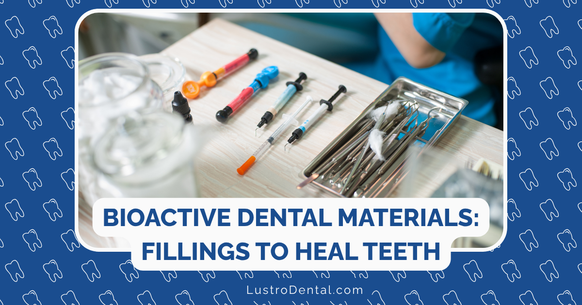
Bioinks for Dental Applications
The choice of bioink is crucial for successful dental bioprinting. According to JSC Medical, several types are being investigated:
Natural Hydrogels
- Collagen: Mimics the natural extracellular matrix of many dental tissues
- Alginate: Offers good printability and cell compatibility
- Fibrin: Promotes cell adhesion and tissue development
- Hyaluronic acid: Supports wound healing and tissue regeneration
Synthetic Polymers
- Poly(ethylene glycol) (PEG): Customizable mechanical properties
- Polycaprolactone (PCL): Provides structural support for bone applications
- Poly(lactic-co-glycolic acid) (PLGA): Controlled degradation rate
Composite Bioinks
Most promising for dental applications are composite bioinks that combine:
- Natural hydrogels for cell compatibility
- Synthetic polymers for mechanical strength
- Ceramic particles (for bone applications)
- Growth factors and bioactive molecules
A review in Frontiers in Bioengineering and Biotechnology highlighted that these composite approaches are showing the most promise for replicating the complex structures of dental tissues.
Stem Cells in Dental Bioprinting
Sources of Dental Stem Cells
Dental tissues are rich sources of stem cells with unique regenerative properties. According to PMC, the main types include:
Dental Pulp Stem Cells (DPSCs)
- Source: Pulp of permanent teeth
- Properties: Highly proliferative, can differentiate into odontoblasts, osteoblasts, and neural cells
- Applications: Pulp regeneration, dentin formation, bone regeneration
Stem Cells from Human Exfoliated Deciduous Teeth (SHED)
- Source: Pulp of baby teeth
- Properties: Higher proliferation rate than DPSCs, excellent differentiation potential
- Applications: Pulp regeneration, neural tissue applications
Periodontal Ligament Stem Cells (PDLSCs)
- Source: Periodontal ligament
- Properties: Can differentiate into cementoblasts, osteoblasts, and fibroblasts
- Applications: Periodontal tissue regeneration
Stem Cells from Apical Papilla (SCAPs)
- Source: Developing tooth roots
- Properties: High proliferative capacity, robust differentiation potential
- Applications: Root and pulp regeneration
Dental Follicle Stem Cells (DFSCs)
- Source: Dental follicle surrounding developing teeth
- Properties: Can form cementum, PDL, and alveolar bone
- Applications: Periodontal complex regeneration
Stem Cell Integration in Bioprinting
The process of incorporating stem cells into bioprinted dental constructs involves several critical steps:
- Cell isolation and expansion: Obtaining sufficient quantities of the appropriate stem cells
- Cell encapsulation: Incorporating cells into bioinks while maintaining viability
- Printing parameters: Optimizing pressure, temperature, and speed to minimize cell stress
- Post-printing culture: Providing the right environment for cell differentiation and tissue maturation
Research cited by McKinney Dentist indicates that successful integration of stem cells in bioprinted constructs is showing promising results in preclinical studies, with some approaches now moving toward early clinical trials.
Challenges and Limitations
Despite its enormous potential, dental bioprinting faces several significant challenges:
Technical Challenges
Vascularization
Creating blood vessel networks within bioprinted tissues remains a major hurdle. Without proper vascularization, larger tissue constructs cannot receive adequate oxygen and nutrients, limiting their viability.
Current approaches include:
- Co-printing with endothelial cells
- Creating sacrificial channels that later form vessel networks
- Incorporating angiogenic factors to promote vessel growth
Resolution and Precision
Dental tissues have microscopic features that are challenging to replicate with current bioprinting technologies. For example, dentinal tubules are approximately 2-3 micrometers in diameter, below the resolution of many bioprinters.
Mechanical Properties
Achieving the appropriate mechanical strength in bioprinted constructs, particularly for hard tissues like dentin and enamel, remains difficult. Current materials often lack the necessary strength and wear resistance.
Biological Challenges
Cell Viability and Functionality
Maintaining high cell viability throughout the printing process and ensuring proper cell function afterward is critical but challenging.
Tissue Maturation
Bioprinted constructs often require extended periods in bioreactors to develop into functional tissues, making the process time-consuming.
Immune Compatibility
Even when using autologous cells (from the patient), the materials and processing involved in bioprinting can potentially trigger immune responses.
Regulatory and Practical Challenges
Regulatory Approval
As noted in PMC, bioprinted dental constructs face complex regulatory pathways, with uncertainties about how they will be classified and evaluated by agencies like the FDA.
Cost and Accessibility
Current bioprinting technologies are expensive and require specialized expertise, limiting their accessibility to specialized research centers.
Scalability
Scaling bioprinting from individual, custom applications to widespread clinical use presents significant challenges in standardization and quality control.
The Future of Dental Bioprinting: What’s on the Horizon?
Despite these challenges, the field is advancing rapidly. Here’s what experts predict we might see in the coming years:
Near-Term Developments (2025-2030)
Clinical Trials for Periodontal Applications
According to PMC, clinical trials evaluating bioprinted constructs for periodontal defects are already showing promising outcomes in tissue integration and regeneration. By 2030, we may see FDA-approved bioprinted products for specific periodontal applications.
Advanced Bioinks
Researchers are developing “smart” bioinks that respond to environmental cues, release growth factors on demand, and better mimic the extracellular matrix of dental tissues.
Improved Vascularization Strategies
New approaches to creating vascularized constructs, such as pre-vascularization techniques and hybrid bioprinting methods, are likely to overcome current limitations in creating larger, more complex dental tissues.
Medium-Term Possibilities (2030-2035)
Functional Pulp Regeneration
Complete, functional regeneration of dental pulp tissue using bioprinting may become clinically available, potentially transforming endodontic treatment.
Partial Tooth Structures
Bioprinted dentin-pulp complexes or root structures might be used in conjunction with traditional materials for hybrid restorations.
Chairside Bioprinting
Simplified bioprinting systems might begin to appear in specialized dental practices, allowing for same-day bioprinting of certain tissue constructs.
Long-Term Vision (Beyond 2035)
Complete Tooth Regeneration
As suggested by research from Essential Endodontics, the ultimate goal of bioprinting entire functional teeth may become reality, potentially eliminating the need for traditional dental implants.
Personalized Regenerative Treatments
Treatments could be tailored to individual patients based on their genetic profile, stem cell characteristics, and specific tissue needs.
Integration with Other Technologies
Bioprinting is likely to be combined with other emerging technologies such as gene editing, nanotechnology, and artificial intelligence to create more sophisticated regenerative solutions.
Implications for Dental Practice and Patient Care
How Bioprinting Could Transform Dental Treatment
If bioprinting fulfills its promise, dental practice could change dramatically:
Shift from Replacement to Regeneration
Rather than replacing damaged or missing tissues with artificial materials, dentistry would focus on regenerating natural tissues.
Personalized Treatment Planning
Digital scans would be used to create precisely customized tissue constructs for each patient’s unique anatomy and needs.
Minimally Invasive Approaches
With the ability to regenerate tissues, more conservative approaches to preserving natural tooth structure would become possible.
Patient Benefits
The potential benefits for patients are substantial:
Biological Integration
Bioprinted tissues would integrate naturally with the body, potentially lasting a lifetime.
Reduced Complications
Natural tissues would avoid many of the complications associated with artificial materials, such as wear, fracture, or rejection.
Improved Aesthetics and Function
Regenerated natural tissues would likely provide better aesthetics and function than artificial replacements.
Long-Term Health Benefits
Preserving or regenerating natural tissues could have positive effects on overall oral health and systemic health.
Conclusion: Preparing for a Regenerative Future
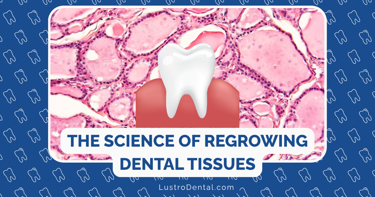
Bioprinting represents a paradigm shift in how we approach dental tissue regeneration. While significant challenges remain, the rapid pace of advancement suggests that bioprinted dental tissues may become a clinical reality sooner than many expect.
For dental professionals, staying informed about these developments will be crucial. The integration of bioprinting into dental practice will require new skills, knowledge, and approaches to treatment planning.
For patients, these advancements offer hope for more natural, long-lasting solutions to common dental problems. While complete tooth regeneration may still be years away, incremental advances in tissue regeneration are already beginning to change what’s possible in dental care.
The journey from today’s experimental bioprinting to tomorrow’s routine regenerative treatments will be fascinating to watch—and participate in. As this technology continues to evolve, it promises to transform not just how we treat dental conditions, but how we think about the very nature of dental health and restoration.
What aspects of dental bioprinting are you most excited about? Do you have questions about how this technology might impact specific dental conditions? Share your thoughts in the comments below!